HCM Resources & Support
Understanding Cardiomyopathy -
A Starter's Guide
A clear, visual guide to the changes that can happen inside the heart with hypertrophic cardiomyopathy.
Created for patients by patients because we believe patients can and should understand more. Offering insight into what is happening in an HCM heart.
HCM Is More Than A Thickened Heart Muscle
Hypertrophic cardiomyopathy can affect the organization of the heart muscle cells, the amount of blood the heart can hold, the heart’s ability to relax, and the electrical system that controls the heartbeat.
Breaking Down The Name
hypertrophic cardiomyopathy
hyper = excessive
trophic = growth
cardio = heart
myo = muscle
pathy = disease
HCM & The Chambers Of The Heart
Let’s talk heart basics and go from there to understand HCM basics.
The heart is an organ that pumps blood throughout your body.
The heart pumps blood throughout the body by contracting and releasing. The blood travels through one-way valves around the heart and to and from the body.
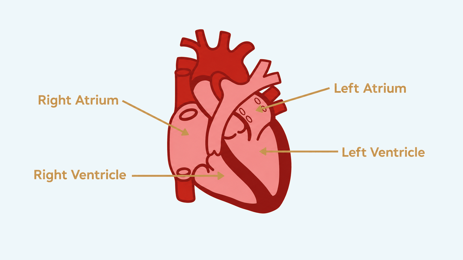
The heart has four chambers. Two upper, or atria, that receive blood. Two lower, or ventricles, which are stronger and pump blood out.
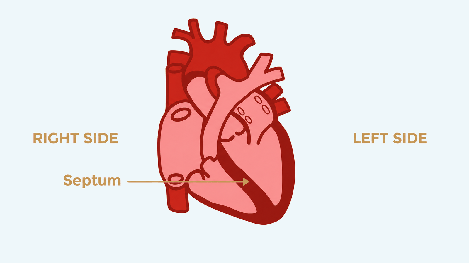
The heart has a right and a left side. The septum is the wall separating the sides of the heart. On each side, there is an atrium and a ventricle.
The right side handles deoxygenated blood, meaning the blood which has circulated the body and distributed oxygen and now needs to get to the lungs to be oxygenated.
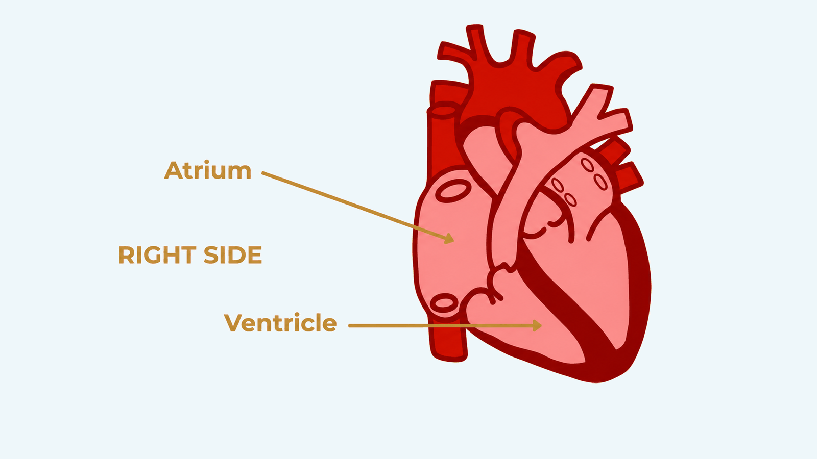
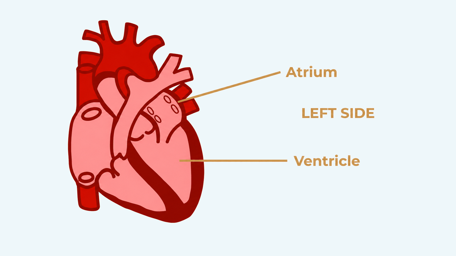
The left side handles the oxygenated blood from the lungs, coming in and being pumped out to the body.
Valves control the blood flow between these chambers by opening and closing and should keep blood moving in the right direction.
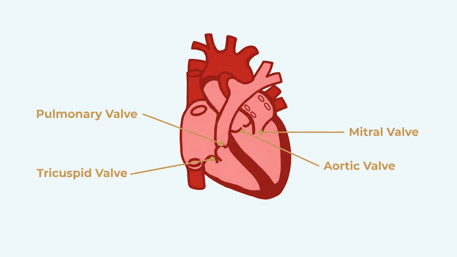
These are the four chambers and valves.
We’ll start with the right atrium. It’s on the right, so we know it handles deoxygenated blood. Being the atrium, we know it receives blood.
So the right atrium receives oxygen-poor blood from the body.
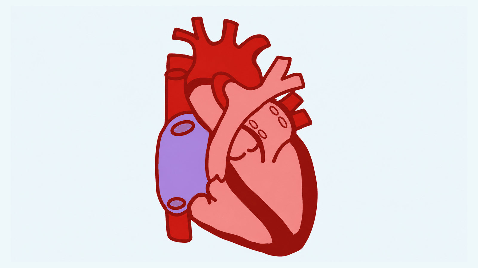
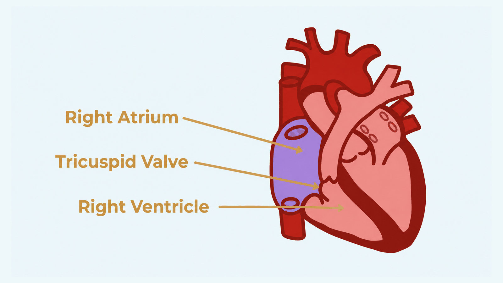
The blood then moves through the tricuspid valve to the right ventricle.
This is the right ventricle. Being on the right, we know it still handles deoxygenated blood. As a ventricle, we know it pumps blood. The right ventricle pumps deoxygenated blood to the lungs.
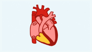
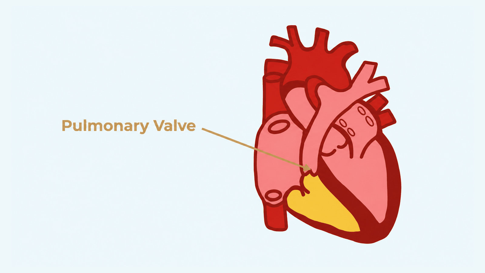
The blood travels through the pulmonary valve into the pulmonary artery, which carries it to the lungs to receive oxygen.
In the lungs, the blood is oxygenated. It then comes back to the heart.
Since the atria collect and the left side handles oxygenated blood, it will move into the left atrium.
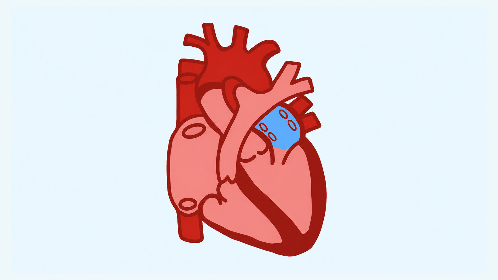
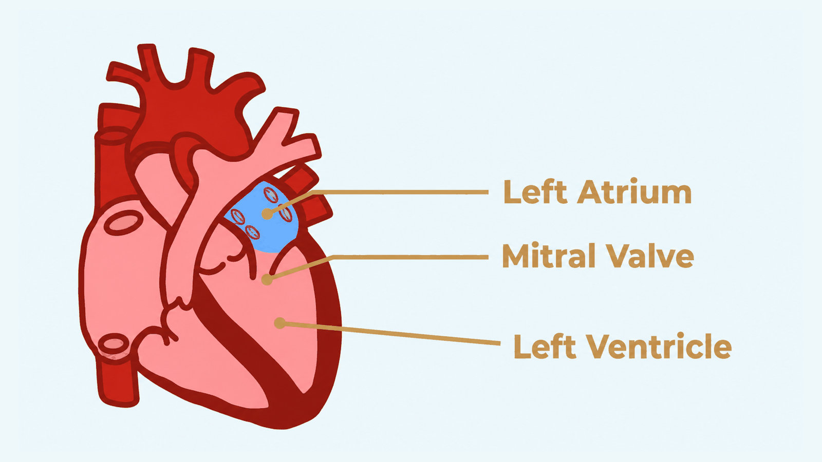
The left atrium receives the oxygen-rich blood from the lungs. It then sends that blood to the left ventricle through the mitral valve.
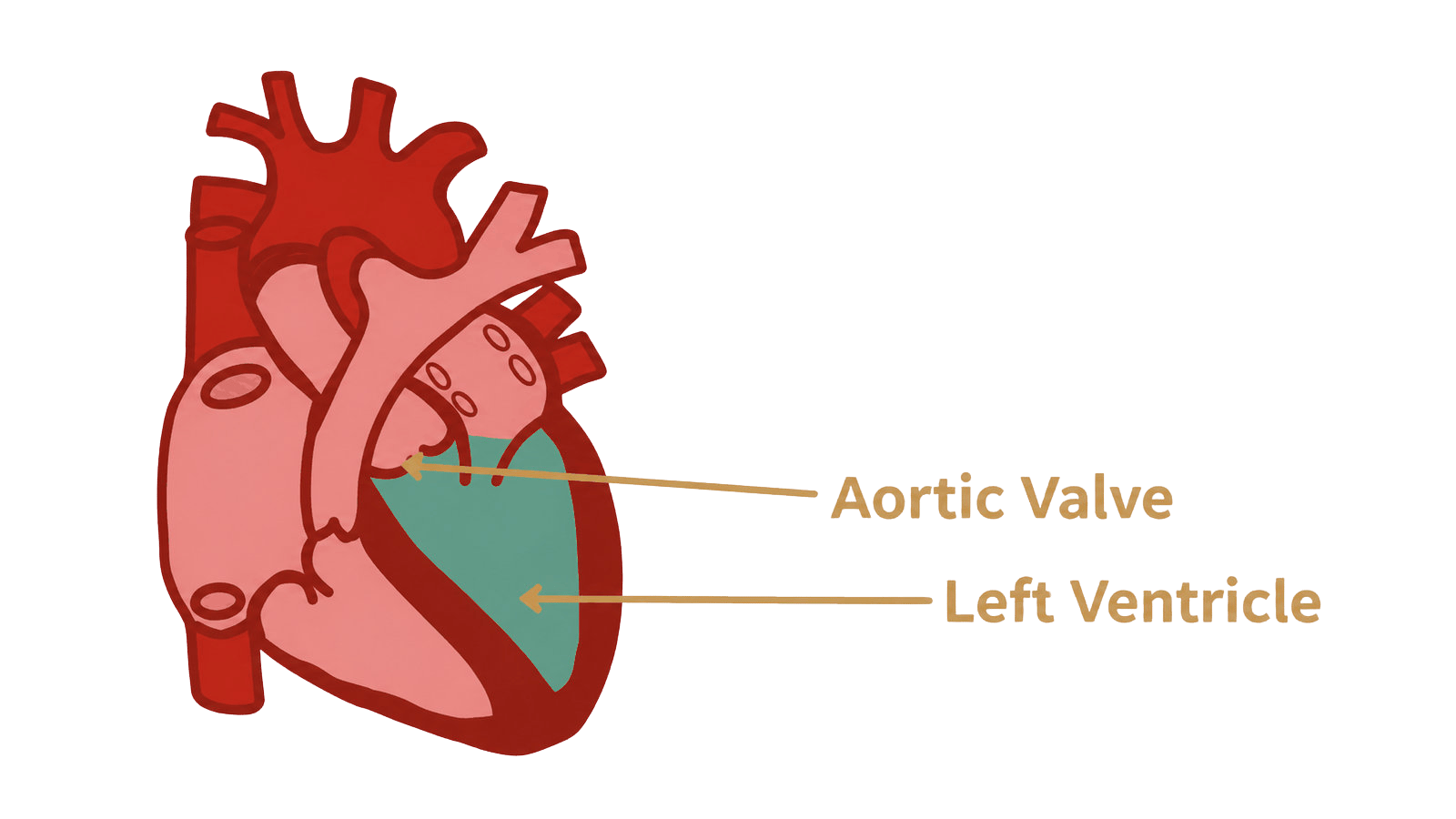
Here is the left ventricle. As a ventricle, it pumps out blood. Being on the left, it handles oxygenated blood. So the left ventricle pumps oxygenated blood out to the body.
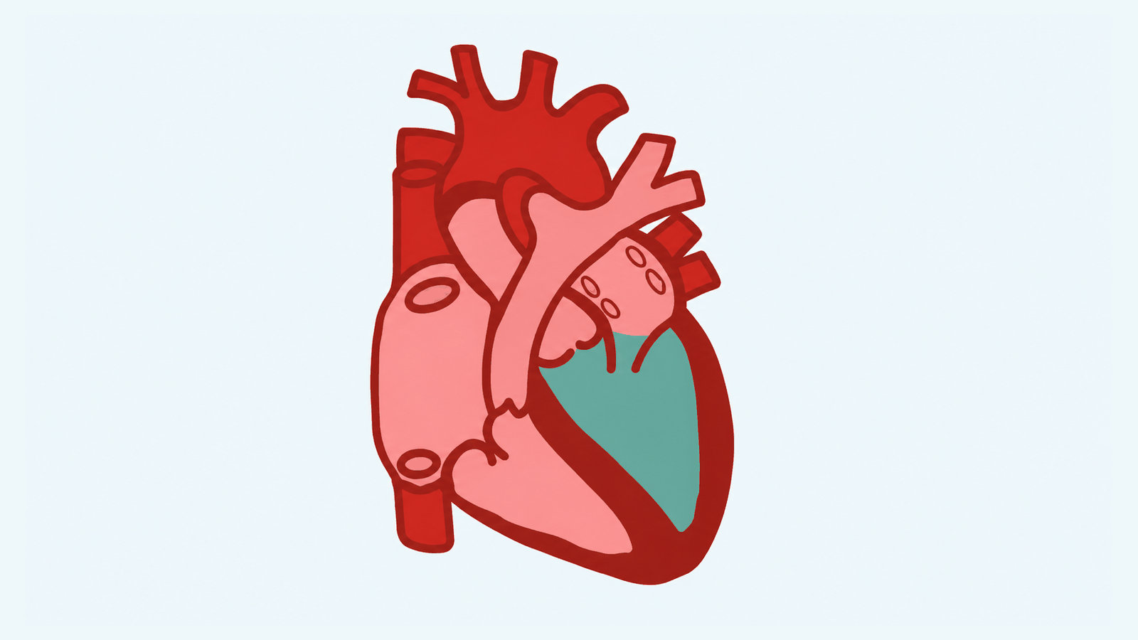
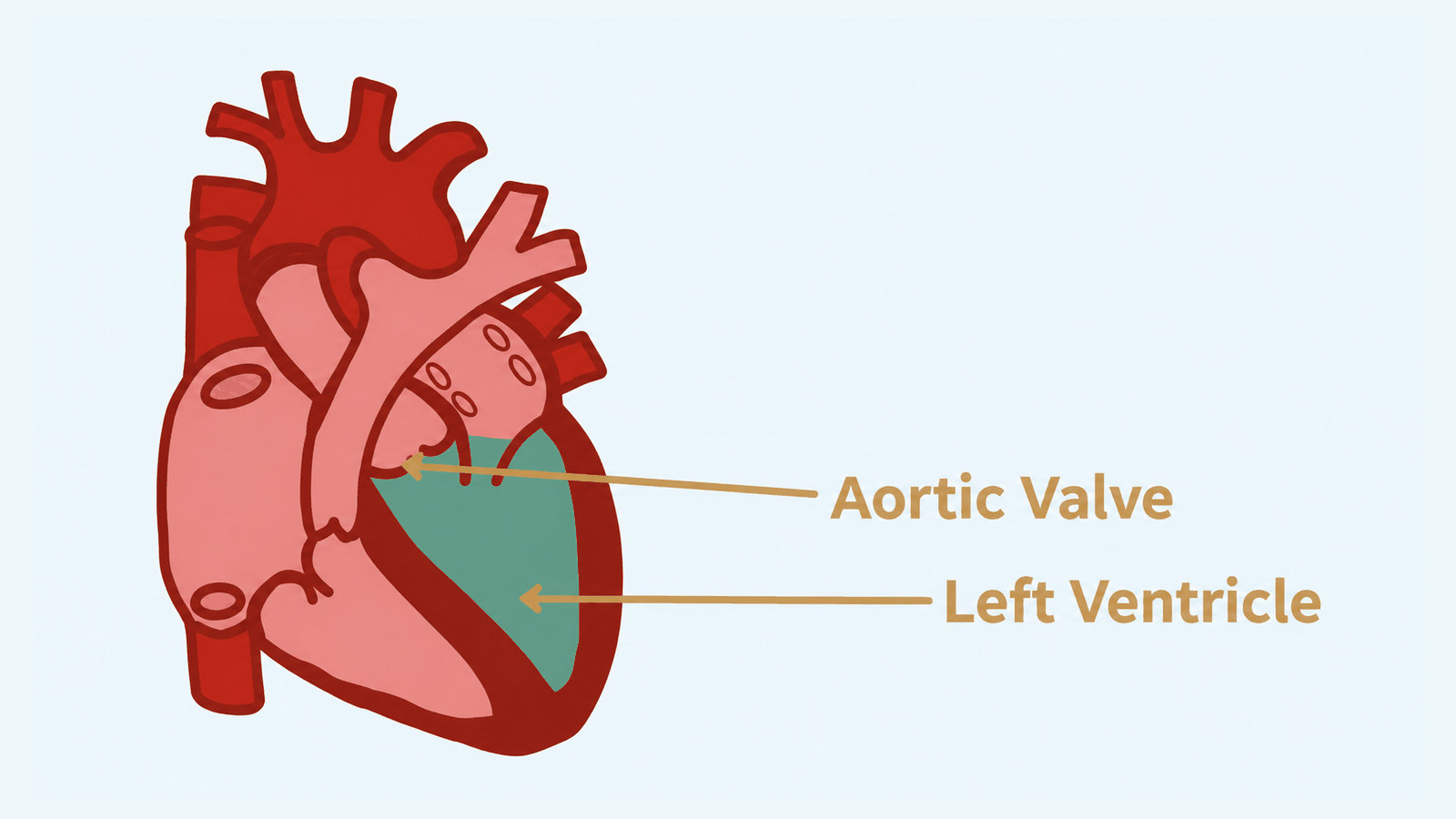
The left ventricle pumps oxygenated blood out to the body through the aortic valve to the aorta, which carries the blood to the body’s tissues and organs. That pumping is key to the heart’s job of circulating blood.
Contracting and relaxing cause the pumping action. Contracting pumps blood out. Relaxing allows the chambers to refill. Valves closing makes sure there is no backflow.
We find HCM primarily in the left ventricle, which should be the heart’s strongest chamber.
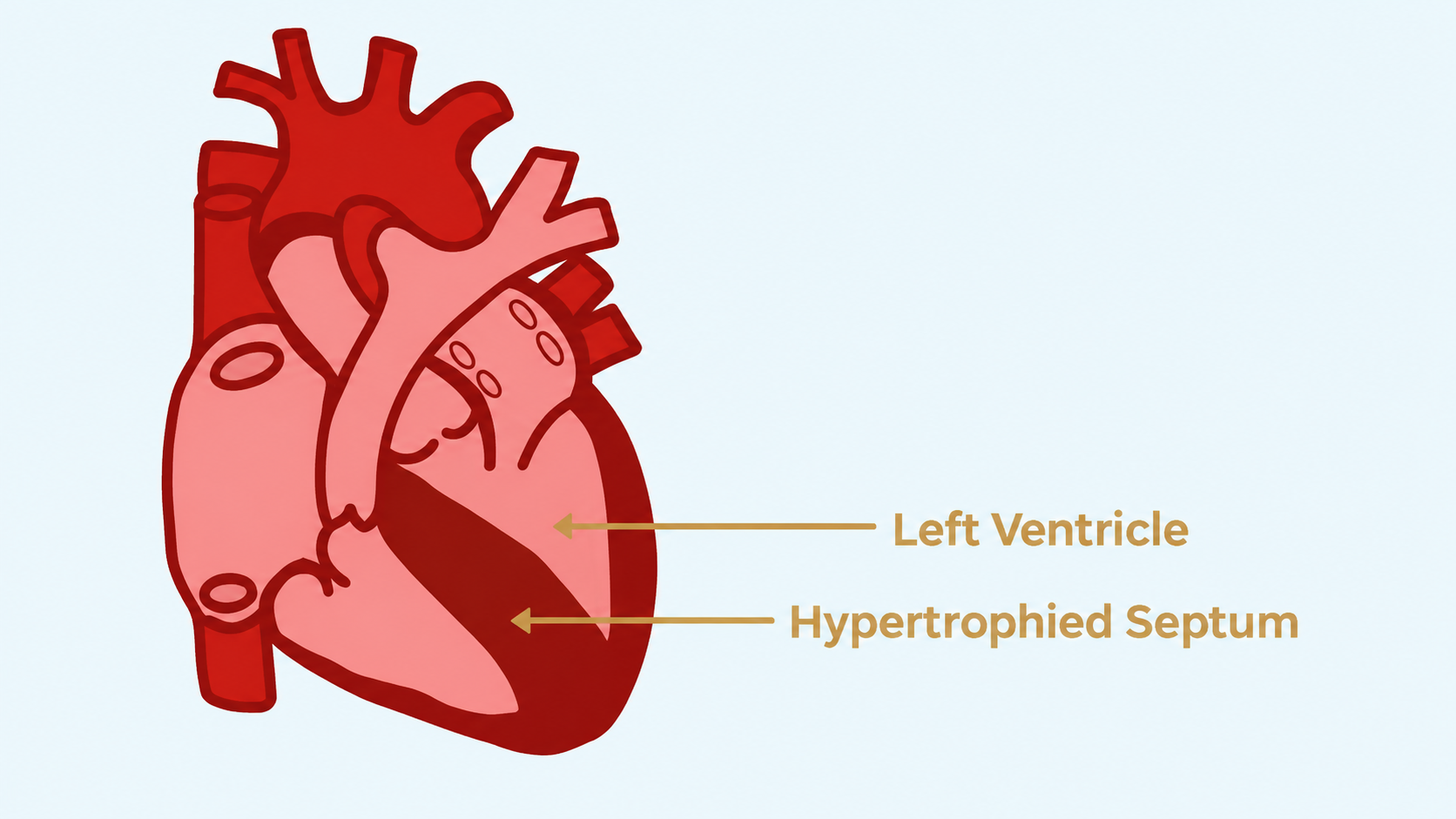
It is where the heart receives oxygen-rich blood and then forcefully pumps it out to the entire body.
Knowing that can help us understand how hypertrophic cardiomyopathy interferes with the functioning of the heart.
HCM Is...
chaotic | chaotic dissarray
extra | extra work
more | more growth
less | less blood
disruption | electrical disruption
rigid | rigid not relaxed
HCM is Chaos And Disorganization Of The Fibers Of The Heart.
This can cause irregular heartbeats and impaired pumping.
An HCM heart will have myocardial disarray. That means the myocyte, or muscle cells, are not organized and aligned in a parallel manner like in a healthy heart. Instead, there is chaos and a decrease in parallel patterns.
This can be described as architectural disorganization. The fibers can be jumbled, have different orientations, and form bizarre bundles.
The genetic mutation in HCM triggers these changes. This may be seen even before any hypertrophy or thickening. Then scarring can add to the disruption.
The disorganized structure leads to inefficient contractions and electrical instability, which cause irregular heartbeats and impaired pumping efficiency.
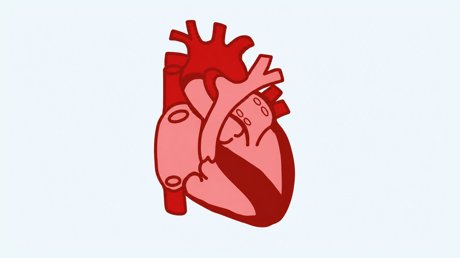
HCM is Extra Work By The Heart Cells
The hyperdynamic heart may lead to heart failure.
From the left ventricle, blood is pumped out to the body. An efficient heart pumps out about 55–65% of the blood in the ventricle with each beat.
With HCM, the heart is often constantly pumping out a higher percentage of blood, in the 65–70–75% range.
The HCM heart is hyperdynamic. The amount of activity is abnormal. The heart is contracting more forcefully.
The cells are doing extra work, squeezing more than they should need to. On top of that, they are not relaxing as much as they should.
Working harder does not mean the heart is working more effectively; in fact, it often results in the opposite, becoming ineffective.
Despite the high contractile force, the heart may still not meet the body’s metabolic demands and heart failure can result.
HCM Is More Growth of the Heart Wall
This can lead to inefficient filling and pumping of the heart.
One way hypertrophic cardiomyopathy affects the heart is with thickened walls of the heart muscle.
This is hypertrophy which occurs outside of other causes, such as high blood pressure, chronic kidney disease, infiltrative diseases, etc.
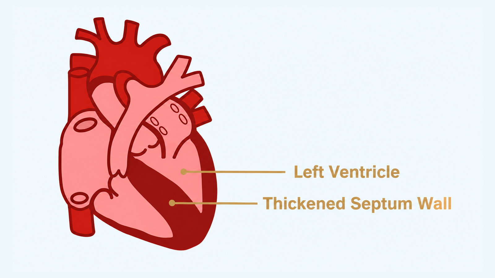
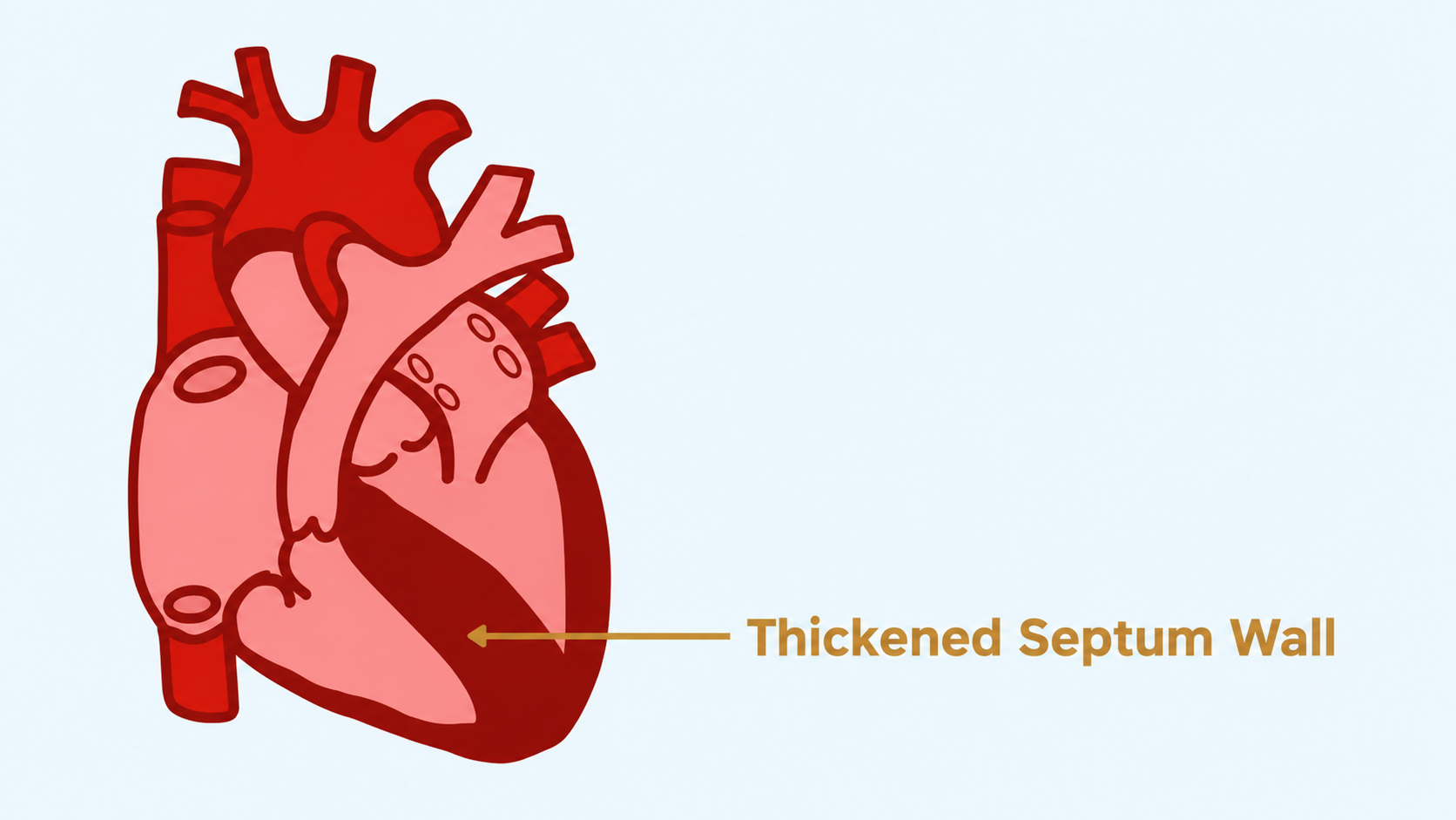
Generally, it is the ventricular septum, or wall between the left and right ventricles, that becomes thickened. It could be the apex or free, outside wall of the left ventricle as well.
In HCM, the heart usually undergoes a period of growth or hypertrophy before staying around similar thickness. An echocardiogram, or echo, can show the thickness.
Normal adult heart walls are usually 10–12 mm thick. The generally accepted cut-off for excessive hypertrophy is 15 mm, or 13 mm with a family history or in someone who is gene positive.
For a child or someone with a smaller heart, this measurement would be dependent on the typical heart size for someone of their body size.
HCM Is Less Room For Blood In The Left Ventricle
The result can be shortness of breath, fatigue, and heart failure.
Hypertrophic cardiomyopathy affects the pumping of the heart. A thickened wall takes up more space and leaves less space for blood in the left ventricle.
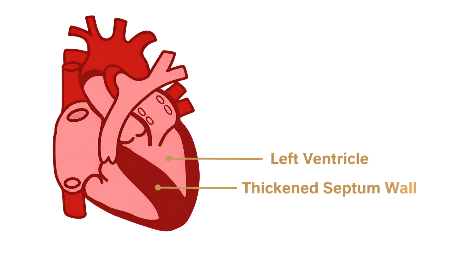
Remember that the heart pumping requires contracting and relaxing, pushing out and refilling. For an adult, the left ventricle holds about 140 mL of blood and ejects 55–65% of that out into the body with each beat.
If the left ventricle is holding less blood, it would need to pump, or eject, a higher percentage, or fraction, of the blood in the left ventricle out with each beat.
This is called a high ejection fraction.
The left ventricle might almost have to empty with each beat, and the heart would struggle to relax and fill properly.
This can cause shortness of breath and fatigue, poor cardiac output and heart failure, and lack of oxygen even though the contractions are good. The heart is working harder but less efficiently.
HCM Causes The Heart To Be Rigid When It Should Be Relaxed And Stiff Where It Should Be Flexible
A chain of reactions including decreased oxygen to heart cells may result.
The HCM heart experiences fibrosis, or fibrotic scarring. This is thickening or scarring of connective tissues, or collagen, that normally happens in response to an injury.
Even though this is a repair response, it becomes detrimental over time. With HCM, the body believes there is an injury and the response of the body makes it real.
The tissue in the heart presenting with HCM is stiffer, thicker, less flexible, and definitely disorganized. It is non-functional and doesn’t contract or conduct electricity like healthy heart muscle.
What problems does this cause?
Both the structure and the electrical function of the heart are affected.
The HCM heart is less efficient. The heart walls don’t relax and expand as they should to fill between beats. This leads to less blood pumping out, which means less oxygen and nutrients getting to the body. Heart failure can occur.
The tissue in the HCM heart does not react or conduct like healthy tissue, so the electrical function of the heart is affected. The fibrotic areas can disrupt the electrical pathways and lead to dangerous arrhythmias and even sudden cardiac death.
An HCM heart may become stiff for several reasons that build upon one another. Gene mutations can cause heart muscle cells to enlarge.
Then muscle walls can become thickened. Additional thickening can also be the result of the heart muscle having to contract more forcefully when pumping blood.
The extra work can also trigger the cells of the heart to enlarge and become disorganized. The abnormal growth then can lead to more scarring and the accompanying stiffness.
Mechanically, the fibrosis can reduce blood flow and oxygen supply to the heart cells. The fibrosis can be seen on a cardiac MRI.
HCM Is Disruption Of The Electrical System Of The Heart
Sudden cardiac arrest can be the result.
HCM can cause electrical problems in the patient’s heart. The myocardial disarray and fibrosis are at the heart of that. Having a thickness of or above 30 mm is a significant risk factor as well.
The fibrosis or scarring makes the heart tissue rigid and stiff instead of like normal heart tissue, which is healthy and elastic.
The scar tissue disrupts the electrical pathways of the heart, which can lead to irregular heartbeats, or arrhythmias, and even sudden cardiac death.
The degree of left ventricular outflow tract obstruction can also increase the risk of sudden cardiac death and is used in the HCM sudden cardiac death risk calculator.
Apical aneurysms, a topic for another time, which can occur with HCM also increase risk.
Disclaimer: This content is not intended to be a substitute for receiving professional medical advice, diagnosis, or treatment. This is a guide to help educate patients, and caregivers, so they may be informed partners in their healthcare.